Product Details

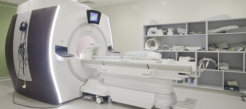
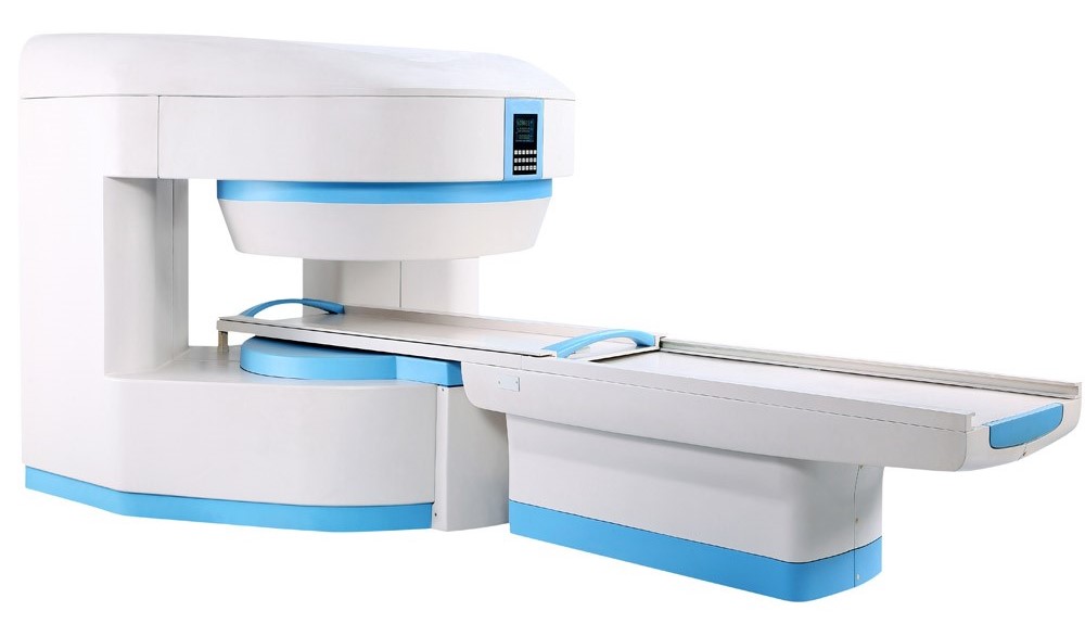
Open & closed Magnetic Resonance Imaging (MRI) Scanner
A magnetic resonance imaging system with permanent magnet field strength, leading hardware preparation configration, supplemented with human space design and advanced matured software system, make the patient more comfortable during the MRI examination process, warm, and to offer doctors accurate results with perfect MRI images to help diagnosis.
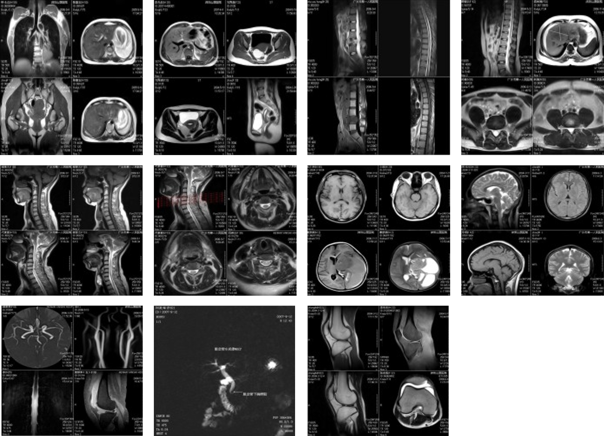
This machien helps a doctor diagnose a disease or injury, and it can monitor how well you’re doing with a treatment. MRIs can be done on different parts of your body.
An MRI of the brain and spinal cord looks for:
- Blood vessel damage
- Brain injury
- Cancer
- Multiple sclerosis
- Spinal cord injuries
- Stroke
An MRI of the heart and blood vessels looks for:
- Blocked blood vessels
- Damage caused by a heart attack
- Heart disease
- Problems with the structure of the heart
An MRI of the bones and joints looks for:
- Bone infections
- Cancer
- Damage to joints
- Disc problems in the spine
MRI can also be done to check the health of these organs:
- Breasts (women)
- Liver
- Kidneys
- Ovaries (women)
- Pancreas
- Prostate (men)
MRI safty:
- Radiation amount of MRI is only one in ten thousand of X-ray.
- 20 years clinical application of MRI approves it has no harm to human body.
- More people can have examinations: Gravida / fetus, children, reexaminiation and physical examination.
- MRI interventional therapy, harmless to both patients and doctors.
- Advantages of Clinical Application: Whole body examination is available
- Harmless in accordance with the healthy idea of patients
- Higher diagnostic level, decrease the rate of missed diagnosis
- More accurate diagnosis, decrease misdiagnosis.
- More defined informaiton
- Improve the judgement accuracy.
- Help to make better treatment plan.
- More objetive evidence-reduce legal risk
Advanced Performance:
- Powerful image processing technology, powerful industrial computer (lPC) workstation, very convenient for image shearing, processing and storage, etc.
- Better reconstruction technology, adopt seniorreconstruction technologies like iteration/ interpolation, complete the signal data collection and K space filling in extremely short time, gain higher resolution images with much clearer fine structures.
- Software has complete and professional sequences which meet the scanning requirements of different parts and can simply gain high quality images based on its rich clinical experience.
- lndependent-developed operation system, doctors can easily and quickly register the patient, browse image, do advanced post-processing, external device connectivity and other functions.
- Perfect MRl system, greatly improved image quality, greatly shortened scanning time, Rich clinical diagnostic imaging sequences, allow the ,users to obtain the images with highest quality, provide the advanced imaging functions and convenient& powerful image post- processing technology to meet the needs of different customers.
- High-performance shielding room design, professional construction team, ensure the scanning environment, undisturbed from outside, provide excellent scanning and operational environment.
Features:
- Excellent price performance ratio.
- Various sequences, Various image typs, high resolution
- Magnet technology: the open & closed magnet, field strength 0.50-3.0T.
- RF system: full-digital.
- Comprehensive application suite and powerful software included instandard configuration
- Patient friendly appearance
- Minimal sitting requirements
- Low operating Costs
- Permanent magnet – no helium
- Higher revenues; Attractive to more patients and referring physicians
- Excellent Return On-Investment
- Decreased costs-optimized profitability
- Software technology:
- easy printing and network transmission for images;
- Fast automatic correction of a sequence and queuing and scanning functions;
- Any directions, at any angle of three-dimensional imaging;
- Reporting module easy using for MRI diagnosis;
- plenty of MRI imaging sequence.
Gradient System
- Active shield full body gradient coil systems, noise reduction & mute technology
RF System
- Spectrometer:All-digital
- Receiving coils:Built-in preamplifier phased array coil
- Receiving coil type (standard):head, neck, body, knee, spine, shoulders, Breast coil
Pulse Sequence
- Standard IR
- Spin Echo ( SE 2D/3D)
- Multi-slice multi-echo (MSME)
- Gradient Echo (GRE 2D/3D)
- Steady state process gradient echo( SSPGRE)
- Fast spin echo (FSE)
- Single shot fast spin echo(SSFSE)
- Multi shot fast spin echo (MSFSE)
- Inversion recovery (IR)
- Inversion recovery fast spin echo (IRFSE)
- Short time inversion recovery (STIR)
- Fluid attenuated inversion recovery ( FLAIR)
- MRM ,MRU ,MRCP
- TOF 2D/3D MRA
- Diffusion weighted imaging (DWI)
- Echo planar imaging(EPI)
Image
- Acquisition matrix 64/128/256/512
- Resolution 1mm(head 24cm FOV 256X256)
- 1.5MM(body 30cm FOV 256X256)
- 0.5mm(head 24cm FOV 512X512)
- 0.75mm( body 30cm FOV 512X512)
- Maximum display matrix 1024x1024
- FOV 20 ~ 400mm
- Slice Orientation Sagittal, coronal, transversal, any angle any oblique
- Image Type T1 Weighted imaging , T2 weighted imaging ,T2* weighted imaging , proton density imaging;Water Suppressed Imange,Fat Suppressed Image, MRM, MRU,MRCP; Magnetic Resonance angiography (MRA), Diffusion Weighted Imaging (DWI)
Workstation
- New communication user software interface, intuitive and easy to learn, bilingual selection
- The main screen displays: Screen monitoring device, machine monitoring, patient monitoring, multi-parameter monitor the amount of concentrated, high security.
- Machine control parameters: pressure, level, temperature
- Patient monitoring parameters: electro cardio, respiratory, RF
- System operating modes: normal operation mode, a controlled operating mode
- Image processing and analysis: image enhancement, zoom, pan, crop, negatives, window width and window level adjustments, mark or clear text, distance measurement, regions of interest selected, the average pixel value, standard deviation of pixel values, signal values distribution, projection reconstruction
- Network components: DICOM 3.0 standard interface, through the local Ethernet network easily to link camera, diagnosis and treatment workstations, medical information systems, remote diagnostics system and laser printer interface.